THE
BY
M.D., D.D.S.
PROFESSOR OF THE PRINCIPLES AND PRACTICE OF DENTAL SURGERY IN THE BALTIMORE
MEMBER OF THE AMERICAN MEDICAL ASSOCIATION
AUTHOR
OF DICTIONARY OF MEDICAL TERMINOLOGY, DENTAL SURGERY AND THE
SIXTH EDITION:
WITH
1855.
Chapin Aaron Harris (1806-1860)
| 原文 | 翻訳 |
|
CHAPTER TENTH.
IRREGULARITY OF THE TEETH. THE
temporary teeth seldom deviate from their proper place in the alveolar
arch, but with the permanent teeth, irregularity of arrangement is of
frequent occurrence. The incisors and cuspidati are more liable to take
an improper direction in their growth than any of the other teeth. The
first and second molars rarely deviate from their proper place; for,
like the teeth of first dentition, they encounter no obstruction in
their growth and eruption. The
first molars being the first of the second set to appear, the ten
anterior teeth are limited to that part of the arch occupied by the
first set, and if this is too small to admit them, irregularity must of
necessity ensue. The
dentes sapientiæ are sometimes prevented from coming out in the proper
place in the lower jaw, by want of room between the second molars and
the coronoid processes; and in the upper jaw, by a want of space between
the last named teeth and the angle of the jaw. When
a bicuspis is forced from its proper place, it turns inwards towards the
tongue, or outwards towards the cheek, according as it is in the upper
or lower jaw. The cuspidati when prevented from coming out in their
proper place, make their appearance either before or behind the other
teeth. When they come out anteriorly, and they do so more frequently
than posteriorly, they often become a source of annoyance to the upper
lip, excoriating its lining membrane, and sometimes causing ulceration. The
incisors of the upper jaw present a greater variety in the manner of
their arrangement than any of the other teeth. The centrals sometimes
come out before and sometimes behind the arch; at other times, their
sides, next to the median line, are turned either directly or obliquely
forwards towards the lip. The laterals sometimes appear half an inch
behind the arch, looking towards the roof of the mouth; at other times,
they come out in front of the arch, and, at other times again, they are
turned obliquely or transversly across it. When
any of the upper incisors are very much inclined towards the interior of
the mouth, the lower teeth, at each occlusion of the jaws, shut before
them, and become an obstacle to their adjustment. This is one of the
most difficult kinds of irregularity to remedy, and one that often
interferes with the lateral motions of the jaw. The
lower incisors sometimes shut in this manner even when there is no
deviation of the upper teeth to the interior. In this case, the
irregularity is owing to a preternatural elongation of the lower jaw,
which more frequently results from some fault of dentition, than from
any congenital defect in the jaw itself.
Sometimes, the superior maxillary arch is so much contracted, and the
front teeth in consequence so much projected, that the upper lip is
prevented from covering them. Cases of this kind, however, are rarely
met with, but when they do occur, it occasions much deformity of the
face, and forms a species of irregularity very difficult to remedy. From
the same cause, the lateral incisors are sometimes forced from the arch,
and appear behind the centrals and cuspidati, the dental circle being
filled with the other teeth.
There are many other deviations in the arrangement of these teeth. Mr.
Fox mentions one that was caused by the presence of two supernumerary
teeth of a conical form, that came up partly behind and partly between
the central incisors, which, in consequence, were thrown forward, while
the laterals were placed in a line with the supernumeraries; the central
incisors, though half an inch apart, formed one row, and the laterals
and supernumeraries, another. Mr. F. says he has seen three cases of
this kind. This
description of irregularity is rarely met with: the author has, however,
in the course of his practice, met several examples.
p.152 M.
Delabarre says, that cases of a transposition of the germs of the teeth
are occasionally met with, so that a lateral incisor takes the place of
a central, and a central the place of the lateral. A similar
transposition of a cuspidatus and a lateral incisor also, sometimes
occurs. Two cases of this sort have fallen under the observation of the
author. The
incisors of the lower jaw, being smaller than those of the upper, and,
in other respects, less conspicuous, do not so plainly show an
irregularity in their arrangement, nor is the appearance of an
individual so much affected by it. Still it should be guarded against,
for any such disturbance, whether in the upper or lower jaw, is
productive of injury to the health of the teeth, and to the beauty of
the mouth. The
growth of the inferior permanent incisors is sometimes more rapid than
the destruction of the roots of the corresponding temporaries. In this
case, the former emerge from the gums behind the latter, and sometimes
so far back, as very greatly to annoy the tongue, and interfere with
enunciation. At other times, the permanent centrals are prevented from
assuming their proper place, because the space left for them by the
temporaries, is not sufficient for their reception. The irregularity in
the former of these two cases, is greater than in the latter. The same
causes, in like manner affect the laterals. M.
Delabarre mentions a defect in the natural conformation of the jaws, by
which the upper temporary incisors on one side of the median line are
thrown on the outside of the lower teeth, while the corresponding teeth,
on the other side of the same line, fall within. The same disposition,
he says, may be expected, unless the defect is previously remedied,
after the dentition of the permanent teeth.* The author has never met
with more than two cases of this sort, and he did not see the subjects
of even these until after they had become adults.
- - - - -
*Enfin il y a une espèce
de torsion de l'une ou de l'autre mâchoire, et quelquefois de toute les
deux, qui fait que les dents temporaries supérieures antérieures
recouvrent les inférieures, d'apaés la meilleure disposition; tandis
qu'à commencer de la linge médiane, les semblables dents de l'autre
côte, rentrent en dedans des inférieures; il est probable, dans ce cas,
que si l'on n'y obvie, le même disposition se reproduira pour la seconde
dentition.-Traité de la Seconde Dentition, p. 136.
p.153
TREATMENT.
The
means employed in the treatment of irregularity should accord with the
indications of nature. When the irregularity is neither great nor
complicated, and its causes are removed before the nineteenth or
twentieth year, the teeth, without any aid from art, will soon acquire
their proper position.
When, however, the exertions of the economy are unavailing, recourse
should be had to the dentist, who can, in almost every case, produce
symmetry and regularity from deformity. The
practicability of altering the position of a tooth, after the completion
of its growth, was well known to many of the early practitioners, but as
before the commencement of the present century, the more particular
object of the dentist, was, the insertion of artificial teeth, this
branch of dentistry met with little attention. Fauchard and Bourdet were
among the first who turned their attention to orthodontia. They invented
a variety of fixtures for adjusting such of the teeth as were not
rightly arranged; but most of these were so awkward in their
construction, and occasioned so much inconvenience to the patient, that
they were seldom employed. Mr.
Fox was among the first to give explicit directions for remedying
irregularity of the teeth. These have formed the basis of the
established practice for more than forty years, and this long trial has
proved them to be founded upon a knowledge of the laws of the economy,
and much practical experience. In
describing the treatment of irregularity, we shall notice the means by
which some of its principal varieties may be removed; otherwise, the
application of the principles of treatment would not be well understood,
since it must be varied to suit each individual case. As a
general rule, the sooner an irregularity in the arrangement of the teeth
is remedied the better, for the longer a tooth is allowed to occupy a
wrong position, the more difficult will be its adjustment. The position
of a tooth may sometimes be altered, after the eighteenth, twentieth or
even the twenty-fifth year, but, generally, it is better not to delay
the application of the proper means until so late a period; for a change
of this kind may be much more easily effected before the several parts
of the osseous system have acquired their full size, and while the
process of new formation is in vigorous operation, than at a later
period of life. The
age of the subject, therefore, should always govern the practitioner in
forming a prognosis of the practicability of removing an irregularity.
Previously to the eighteenth year, he may almost always form a favorable
one, but after this time, his efforts will be less likely to succeed. The
first thing claiming attention in the treatment of irregularity, is the
removal of its causes. Whenever, therefore, the presence of any of the
temporary teeth has given a false direction to one or more of the
permanent, they should be extracted, and the deviating teeth pressed
several times a day with the finger, in the direction they are to be
moved. This, if the irregularity has been occasioned by the presence of
a deciduous tooth, will, generally, be all that is required. But,
when it is the result of narrowness of the jaw, either natural or
acquired, one of the permanent teeth on each side should be removed, in
order to make room for such as are improperly situated. The second
bicuspids are the teeth which should, generally, be extracted, except
the first permanent molars are so much decayed as to render their
preservation impracticable, or, at least, very doubtful; in which case,
these teeth should be removed in their stead. After the removal of the
second bicuspids, the first, usually, very soon fall back into the
places which they occupied, and furnish ample room for the cuspidati and
incisors. But if the first fail to do this, they may be gradually forced
back by inserting wedges of wood or gum elastic between them and the
cuspidati, or by means of a ligature of silk, or gum elastic, fastened
to the first molar on each side, and securely tied. These should be
renewed, every day, until the desired result is produced. The
most frequent kind of irregularity, resulting from narrowness of the
jaw, is the projection of the cuspidati. These teeth, with the exception
of the second and third molars, are the last of the teeth of second
dentition that are erupted, and are, consequently, more liable to be
forced out of the arch than any of the others, especially when it is so
much contracted as to be almost entirely filled before they make their
appearance. The
common practice, in cases of this sort, is to remove the cuspidati. But
as these teeth contribute more than any of the others, except the
incisors, to the beauty of the mouth, and can, in almost every case, be
brought to their proper place, the practice is injudicious. Instead of
removing them, a bicuspis should be extracted from each side. When the
space between the lateral incisor and first bicuspis is equal to
one-half the width of the crown of the cuspid, the second bicuspis
should be removed, but when it is less, the first should be taken out,
and for the reason, that, although the crown of the latter might be
carried far enough back after the removal of the former, to admit the
crown of the cuspid between it and the lateral incisor, the root of this
tooth would remain in front and partly across the root of the first
bicuspis, leaving a more or less prominent vertical ridge on the
anterior part of the alveolar border, which, to some extent, at least,
would act as an irritant to the gums and periosteum. As
the incisors of the upper jaw are more conspicuous than those of the
lower, and when well arranged contribute more to the beauty of the
mouth, their preservation and regularity are of greater relative
importance. Hence, the removal of a lateral incisor, when it is situated
behind the circle of the other teeth, as is often done, with a view of
remedying the deformity produced by its false position, is a practice
which cannot be too strongly deprecated, provided sufficient space can
be made between the cuspidatus and central incisor for its reception, by
the removal of a bicuspis from each side of the jaw. But,
in describing the treatment of irregularity, we will commence with an
incisor occupying an oblique or transverse position across the alveolar
ridge, so that the cutting edge of the tooth, instead of being in a line
with the arch, forms an angle with it of from forty to ninety degrees.
This variety of deviation is rarely met with in both centrals, but often
occurs with one- Some
dentists have recommended in cases of this sort, when the space between
the adjoining central and lateral incisor is equal to the width of the
deviating tooth, to turn the latter in its socket with a pair of
forceps, or to extract and immediately replace it with the labial face
of the organ outwards. It is scarcely necessary to say, that if a tooth
is extracted or turned in its socket, the vessels and nerves from which
it derives its nourishment and vitality are severed, and though its
connection with the alveolus may be partially re-established, it will be
liable to act as a morbid irritant. FIG.
52.
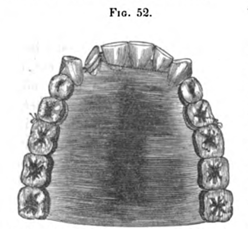
The
tooth, however, may be brought to its proper position without incurring
the risk of injury by accurately fitting a gold ring or band with knobs
on the anterior and posterior edges; to each of these, a ligature should
be attached. The ligatures, thus fastened to the ring, should be carried
back, one on either side, in front and behind the arch, and secured to
the bicuspids in the manner as represented in Fig. 52, so as to act
constantly upon the irregular tooth. These should be renewed from day to
day, until the tooth assumes its proper position. But
before attempting to turn the deviating tooth, it should be ascertained
if the aperture between the adjoining teeth is sufficient to admit of
the operation. If it is not, it should be increased by the extraction of
a bicuspis from each side of the jaw, and moving the teeth in front of
them backwards until sufficient room is obtained. The time required to
do this, will vary from three to eight or ten weeks, depending upon the
number of teeth to be acted on, and the age of the patient.
Narrowness of the alveolar border is sometimes a cause of irregularity
of the upper incisors. In this case, the centrals usually project,
though it sometimes happens that some are in front and some behind the
arch, producing great deformity; to remedy which, the second bicuspids
should be removed, unless the first molars are so much affected by
caries as to render their preservation doubtful. In this event, they
should be extracted, in place of the second bicuspids. The
following case will serve to illustrate the means employed for remedying
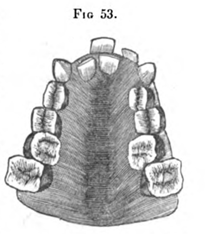
this description of deformity. The subject was a young lady fifteen
years of age, and when placed under the author's care in the fall of
1844, her teeth presented the following arrangement:
The
second molars of the upper jaw occupied their proper position in the
alveolar arch, or in other words, they were a little more than an inch
and a quarter apart; the first molars were hardly an inch apart, and the
first bicuspids were still nearer to each other. The cuspidati, except
having been pushed a little too far forward, occupied, very nearly,
their proper position. The right central and left lateral incisors
projected full a quarter of an inch, lifting and otherwise annoying and
disfiguring the upper lip: the left central was thrown back and partly
between the right central and left lateral, while the right lateral
occupied a position in a line with it.
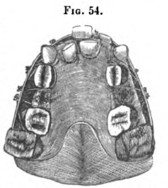
Without going into a minute detail of the method which was adopted for
procuring the appliance employed, it will be sufficient to refer the
reader to Fig. 54. This represents a plaster model of the teeth,
alveolar border, palatine arch, and the apparatus employed for remedying
the deformity. The second bicuspids were first extracted, then, by means
of ligatures applied to the second molars and first bicuspids, and made
fast to a band of gold passing round on the outside of the arch, which
were renewed every day, these teeth were brought out to their proper
position in eleven weeks; this done, there was a space of nearly an
eighth of an inch between the cuspidati and first bicuspids; this was
filled up, by bringing back with ligatures, properly applied, the former
to the latter. A ligature was next applied to the right lateral, passed
through a hole in the gold band in front, and made fast. In ten days
this tooth was brought out to its proper place. A ligature was now
attached to a knob soldered on the gold plate behind the teeth on the
inner side of the alveolar border for the purpose, and tied tightly in
front of the projecting right central incisor. In about three weeks this
was brought to a position along side the lateral incisor of the same
side. The left central was then, in like manner, brought forward and the
left carried backward to its proper place.
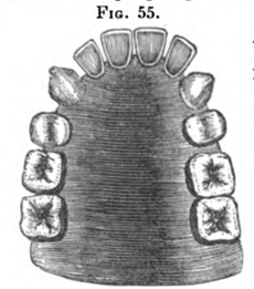
After the deformity was corrected, the teeth presented the arrangement
represented in Fig. 55, taken from a plaster model of the upper jaw. To
correct the irregularity in this case, required, in all, twenty-one
weeks. If all the teeth could have been acted upon at the same time, the
operation might have been accomplished in a shorter period. It was found
necessary, too, in consequence of the diseased action, occasioned by the
apparatus, in the gums, to remove it every eight or ten days, and let it
remain off each time twenty-four hours. It may be proper, also, to
observe, that every time the ligatures were removed, it was taken from
the mouth, and the teeth thoroughly cleansed. For
moving a projecting incisor or cuspidatus backwards, a gold spiral
spring was formerly employed. It was found to be more efficient than a
ligature of silk, inasmuch as it kept up a constant traction upon the
deviating tooth. But it is objectionable on account of the annoyance it
causes the patient. A ligature of gum-elastic is far preferable, and
this material is now very generally employed in the treatment of every
description of irregularity of the teeth in which agencies of this sort
are required.
There are other varieties of irregularity of the upper incisors, but we
shall only notice one, which, from its peculiar character, is sometimes
exceedingly difficult to remedy. It is, when one or more of these teeth
are placed so far back in the jaw, that the under teeth come before them
at each occlusion of the mouth.
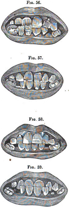
FIG.
56.
Of
this variety, Mr. Fox enumerates four kinds :-The first is, when one of
the central incisors is situated so far back, that the lower teeth shut
over it, while the other central remains in its proper place, as
represented in Fig. 56, copied from his work, as are also those which
follow. FIG.
57. The
second is, when both of the centrals have come out behind the circle of
the other teeth, and the laterals occupy their own proper position, as
represented in Fig. 57. FIG.
58. The
third is, when the lateral incisors are thrown so far back, that the
under teeth shut before them, while the centrals are well arranged, as
exhibited in Fig. 58. FIG.
59. The
fourth kind is, when all the incisors are placed so far behind the arch
that the lower teeth shut before them, as in Fig. 59. He
might also have added to this variety of irregularity, a fifth
description, for it sometimes happens that the cuspidati of the upper
jaw are thrown so far back, as to fall on the inside of the lower teeth.
The author has met with several cases of this description of deviation. Two
things are necessary in the treatment of the kinds of irregularity just
described; the first is, to prevent the upper and lower teeth from
coming entirely together, by placing between them some hard substance,
so that the former may not be prevented by the latter from being brought
forward. The second is, the application of some fixture that will exert
a constant and steady pressure upon the deviating teeth, until they pass
those of the lower jaw. For
the accomplishment of this, various plans have been proposed. Duval
recommends the application of a grooved or guttered plate, and Catalan
has invented an instrument, based, we believe, upon the same principle,
but much better adapted "to the purpose. The inclined plane of Catalan,
although, until we had employed it, we doubted its utility, is certainly
an effectual and speedy method of moving deviating front teeth in the
upper jaw, from behind the dental circle to their proper places. It acts
with great force, and in precisely the proper manner for the
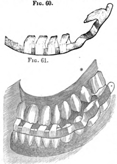
accomplishment of the object. FIG.
60. FIG.
61.
The
accompanying cuts, copied from Catalan, exhibit the manner in which his
inclined plane is constructed. The one here represented, is applied to a
case where all the upper incisors fall behind the lower. Its
construction should be varied to suit the peculiarity of the case. If
but one tooth deviates, only one inclined plane will be required. The
apparatus should also be so adapted and secured to the teeth as to
occasion as little inconvenience to the patient as possible. The
circular bar or plate of gold, running round in front of the teeth,
should reach from the first molar on one side to the first molar on the
other, and the plate, extending up from it should cover the grinding
surfaces of these teeth, and be long enough to cover their lingual
surfaces also, as the whole fixture will thereby be rendered much firmer
and more secure. In
the application of this principle for the correction of irregularity,
the author has been in the habit of constructing the apparatus somewhat
differently. With a brass model and zinc counter-model, he has a plate
of gold struck up over all the teeth, when practicable, as far back as
the first or second molar, so as completely to encase them and the
alveolar ridge in it. An encasement of this sort, possesses greater
stability than can be obtained for an appliance like the one represented
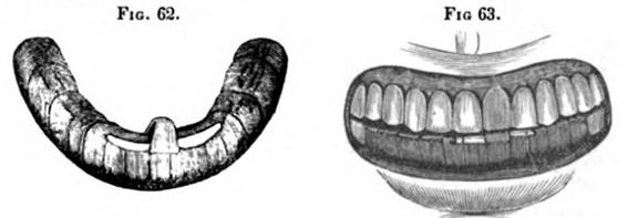
in Figs. 60 and 61. FIG.
62.
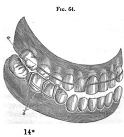
FIG. 64.
In
Fig. 62, is seen a representation of an inclined plane for bringing
forward a central incisor which had come out about a quarter of an inch
behind the circle of the other teeth. The manner of the action of this
instrument upon the deviating tooth is shown in Fig. 63.
p.162 The
plan proposed by Delabarre, is to pass silk ligatures round the teeth,
in such a way that a properly directed and steady pressure will be
exerted on such of the teeth as are situated behind the arch, and to
keep the jaws from coming in contact, he recommends the application of a
metallic grate, fitted to two of the inferior molars, (see Fig. 64,)
taken from his treatise on second dentition. a Represents a ligature
round the teeth, and b a metallic grate on two of the lower teeth. This
plan possesses the merit of simplicity, and occasions but little or no
inconvenience to the patient; but it will sometimes be found not only
inefficient, but also to loosen the teeth adjacent to those to be
brought forward. The force on the irregular teeth, and those against
which the ligatures act, being equal, and in opposite directions, the
latter will be drawn back, while the former are brought forward; and
thus the means used for the correction of one evil, will sometimes
occasion another. FIG.
65.
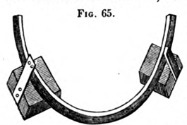
The
means recommended by Mr. Fox, consists of a gold bar about the sixteenth
part of an inch in width, and of proportionate thickness, bent to suit
the curvature of the mouth, and fastened with ligatures to the temporary
molars of each side. It is pierced opposite to each irregular tooth,
with two holes. The teeth of the upper and lower jaw are prevented from
coming entirely together by means of thin blocks of ivory, attached to
each end of the bar by small pieces of gold, and resting upon the
grinding surfaces of the temporary molars. (See Fig. 65.) FIG.
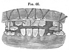
66.
After the instrument has been thus fastened to the teeth, silk ligatures
are passed round such as have deviated to the interior, and through the
holes opposite them, and then tied in a firm knot, on the outside of the
bar. (See Fig. 66.)
p.163 The
ligatures must be renewed every three or four days, until the teeth
shall have come forward far enough to fall plumb on those that formerly
shut before them and acquired a sufficient degree of firmness to prevent
them from returning to their former position. But as soon as the teeth
shut perpendicularly upon each other, the blocks may be removed, and the
bar alone retained.
Since 1830, many practitioners, both in England and the United States
have substituted caps of gold for the blocks of ivory recommended by Mr.
Fox; and, instead of simply bending the bar, they now swage it between a
metallic cast and die, so that all its parts, except those immediately
opposite the irregular teeth, may be perfectly adapted to the dental
circle. The apparatus, with these modifications, is more comfortable,
and less liable to move upon the teeth. Mr.
Fox directs, that the blocks of ivory should be placed upon the
temporary molars, but the caps of gold now substituted are entirely
disconnected from the bar, and are often used after the moulting of
these teeth; they are, therefore, placed upon the first permanent
molars. As
the caps prevent the teeth from coming together, mastication, during the
time they are worn, is, necessarily, performed on them. They should,
therefore, be placed upon the largest and strongest teeth; and it is for
this reason that the molars should be selected. The
curved bar should be washed and the teeth cleansed every time the
ligatures are renewed. If this be neglected, the particles of food that
collect between it and the teeth, will soon become putrid and offensive,
constituting a source of disease both to the gums and teeth. But
before the bar is applied, it should be ascertained whether there is
sufficient space for the reception of the deviating teeth, and if there
is not, room should be made in the manner as before described. Some
diversity of opinion exists as to the most suitable age for the
correction of this description of irregularity. Mr. Fox, it would seem,
preferred the period immediately previous to the moulting of the
temporary molars-probably the tenth or eleventh year after birth. Some
think, that the fore part of the dental arch continues to expand until
the second denture is completed, and that the bicuspids afford a better
support for the ends of the bar than any other teeth, and are,
therefore, content to wait until the fifteenth or even sixteenth year.
But, though the arch does sometimes expand a little, yet even when the
expansion occurs, it is generally so inconsiderable, that very little
advantage can be derived from it. Moreover, the arch, instead of
expanding is much more liable to contract whenever a vacancy occurs in
the dental circle, either by the extraction, or from the improper growth
of one or more of the teeth; hence, the difficulty is apt to be
increased by delay. The
evil, it is true, may be remedied at the fifteenth, seventeenth, or even
eighteenth year; but it is rarely advisable to defer it to so late a
period. The
most that is required in the treatment of irregularity of the lower
incisors, is to remove a tooth, and to apply frequent pressure to the
teeth that are improperly situated. The lower incisors are less
conspicuous than those of the upper jaw, and the loss of one, if the
others are well arranged, is scarcely perceptible.
- - - - - - - - - - - - - -
CHAPTER ELEVENTH.
DEFORMITY RESULTING FROM EXCESSIVE
DEVELOPMENT OF THE
TEETH AND ALVEOLAR
RIDGE OF THE LOWER JAW.
WHEN
the arch described by the teeth of the lower jaw is larger than that of
the upper, the incisors and cuspids of the former shut in front of those
of the latter, causing the chin to project and otherwise impairing the
symmetry of the face. This description of deformity may result from two
causes, namely, excessive development of the teeth and alveolar arch,
and partial luxation and protrusion of the lower jaw. But we shall
confine our remarks in this chapter to the treatment of that produced by
the former.
TREATMENT.
The
remedial indications of the deformity in question, consists in
diminishing the size of the dental arch, which is always a tedious and
difficult operation, requiring a vast amount of patience and
perseverance on the part of the patient, and much mechanical ingenuity
and skill on the part of the dentist. The appliances to be employed
have, of necessity, to be more or less complicated, requiring the most
perfect accuracy of adaptation and neatness of execution; they must also
be worn for a long time, and, as a natural consequence, are a source of
considerable annoyance.
In
the treatment of a case of this sort, the first thing to be done, is to
extract the first bicuspis on each side of the jaw. By this means
sufficient room will be obtained for the contraction, which it will be
necessary to effect in the dental arch, for the accomplishment of the
object. An accurate impression of the teeth and alveolar ridge, should
now be taken, in the manner to be hereafter described, with wax,
previously softened in warm water. From this impression, a plaster model
should next be procured, and afterwards a metallic model and
countermodel.
FIG.
68.
This
done, a gold plate of the ordinary thickness should be swaged to fit the
first and second molars, if the latter has made its appearance, and if
not, the second bicuspis and first molar on each side of the jaw, so as
completely to incase these teeth. If these caps, on applying them to the
teeth in the mouth, should not be found thick enough to prevent the
front teeth from coming together, a piece of gold plate may be soldered
on that part of each which covers the grinding surfaces of the teeth,
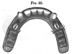
and having proceeded thus far, a small gold knob should be soldered on
each side of each cap, and to each of which a ligature of silk or gum
elastic should be attached. These ligatures should now be brought
forward and tied tightly around the cuspidati. When thus adjusted, the
lower arch will present the appearance exhibited in Fig. 68. By this
means the cuspidati may, in fifteen or twenty days, be taken back to the
bicuspids; but, if in their progress they are not carried towards the
inner part of the alveolar ridge, the outer ligatures may be left off
after a few days, and the inner ones only employed, to complete the
remainder of the operation.
After the positions of the cuspidati have been thus changed, the gold
caps should be removed and a circular bar of gold, extending from one to
the other, so constructed as to pass about a quarter of an inch behind
the incisors, should now be soldered at each end to the inner side of
each cap, and a hole made through it behind each of the incisors,
through which a ligature of silk should be passed, and after it is
placed in the mouth, brought forward and tied tightly in front of each
tooth. These ligatures should be renewed every day until the teeth are
carried far enough back to strike on the inside of the corresponding
teeth in the upper jaw.
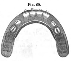
Fig.
69 represents the appearance which a plaster model of the lower jaw
presents with the last named apparatus on it, and an examination of this
will convey a more correct idea of its construction, and the manner of
its application, and mode of action, than any description which can be
given.
FIG.
69.
The
author has never known an apparatus of this description to be employed
by any one but himself; in his practice it has proved more efficient
than any contrivance which he has ever used. But it is necessary that
the caps be fitted with the greatest accuracy to the teeth to which they
are applied, and they should be removed every day and thoroughly
cleansed, as well as the teeth they cover. If this precaution is
neglected, the secretions of the mouth, which collect between the gold
caps and the teeth, will soon become acrid and corrode the latter. This
precaution, therefore, should always be most strictly observed.
p.168 - - - - - - - - - - -
CHAPTER TWELFTH.
PROTRUSION OF THE LOWER JAW. THIS
deformity, although produced by a different cause from the one last
described, is precisely similar to it, and gives to the lower part of
the face an exceedingly morose and disagreeable appearance. It also
interferes with mastication, and often with prehension and distinct
utterance. It wholly changes the relationship which the teeth should
sustain to each other when the mouth is closed. The cusps or
protuberances of the bicuspids and molars of the jaw, instead of fitting
into the depressions of the corresponding teeth of the other, often
strike their most prominent points; at other times the outer
protuberances of the lower molars and bicuspids, instead of fitting into
the depressions of the same class of teeth in the upper jaw, shut on the
outside of these teeth. The trituration of aliments is consequently
rendered more or less imperfect. The
description of deformity under consideration is characterized by the
protrusion of the lower jaw, and in the opinion of Dr. J. S. Gunnell, is
the result of a "natural partial luxation." It is of more frequent
occurrence than the one which results from excessive development of the
teeth and alveolar ridge, and requires, as before stated, an entirely
different plan of treatment. It rarely occurs previously to second
dentition, but, Dr. G. says, he has met with it previously to that
period.
TREATMENT. The
plan of treatment which Dr. Gunnell proposes, and it is unquestionably
the most successful that has ever been employed, consists in fastening a
small block of ivory on one of the lower molars, thick enough to keep
the front teeth about a quarter of an inch apart when the jaws are
closed. He then puts on Fox's bandage, buckles it as tight as the
patient can bear with convenience, pressing "the chin upwards and
backwards." Then, if the teeth be irregular, he takes "a piece of tough
wood of the shape of a narrow spoon-handle," which he introduces between
the teeth and presses on, firmly, the "outside of the front lower
protruding tooth or teeth, and on the inside of the upper irregular
teeth, for from five to ten minutes, two or three times a day; the lower
end of the stick or piece of wood and hand being below the chin, thereby
pressing the lower teeth inwards and backwards, and the upper teeth
outwards and forwards. In this way, says Dr. G., "I have restored the
face or jaws to their proper symmetry in one week, though occasionally
it will take from three to six weeks or even longer." FIG.
70.
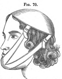
The
description of bandage here alluded to, and the manner of its
application is represented in Fig. 70. When the protrusion of the lower
jaw is accompanied by irregularity, Dr. G. very properly directs that
means should, at the same time, be employed for remedying it. He also
recommends that the operation for retracting the protruding jaw, be
performed as soon as the deformity occurs; though, he says, it may be
successfully remedied at any time. previously to the age of puberty, and
that he has done it at a much later period, but that after the sixteenth
year of age, the operation becomes more difficult. In
cases where the lower front teeth shut over the upper, and thus cause a
deformity of the face, it is important to discriminate correctly between
those which result from malformation, and a protrusion of the jaw
occasioned by partial luxation, as the remedial indications in the two
are entirely different. Those which would prove successful in the one,
would prove unsuccessful in the other. But, fortunately, deformity
arising from this last mentioned cause, is, comparatively, of rare
occurrence; hence the dentist is seldom called upon to exercise his
ingenuity and skill in its treatment.
p.171 - - - - -
CHAPTER THIRTEENTH.
PECULIARITIES IN THE FORMATION AND GROWTH OF
THE TEETH. IN
the development and growth of the various parts of the body, curious and
interesting anomalies are sometimes observed; but in no portion of it,
are they more frequent in their occurrence, or diversified in their
character than in the teeth. But aberrations in the formation and growth
of these organs, are, for the most part, confined to the teeth of second
dentition.
Mr.
Fox gives a drawing of a tooth very nearly resembling the letter S. The
malformation was caused by an obstructing temporary tooth. The author
has also met with several examples of teeth similarly deformed, and from
like causes.
The
molars of the upper jaw sometimes have four and even five roots, and
those of the lower jaw, three and occasionally four. The crowns of the
teeth, also, frequently present deviations from the natural shape
equally striking and remarkable.
The
next peculiarity to be noticed, is that of size, and in this respect the
teeth are very variable. Even in the same mouth, the want of relative
proportion between the different classes of teeth, is sometimes quite
conspicuous. But examples of this kind are not very frequent, for where
there is an increase or diminution in the size of the teeth of one
class, there is generally a corresponding change in those of the other.
Aberrations of this character are probably dependent upon some diathesis
of the general system, whereby the teeth, during the earlier stages of
their formation, are supplied with an excessive or diminished quantity
of nutriment.
Some
very remarkable deviations have been known to take place in the growth
of the teeth. The most singular case on record, is that narrated by
Albinus: "Two teeth," says he, "between the nose and the orbits of the
eye, one on the right side, and the other on the left, were enclosed in
the roots of those processes that extend from the maxillary bones to the
eminences of the nose. They were large, remarkably thick, and so very
like the canini, that they might have seemed to be these teeth
themselves, which had not before appeared; but the canines themselves
were also present, more than usually small and short, and placed in
their proper sockets. The former, therefore, appear to have been the new
canini, which had not. penetrated their sockets, because they were
situated where these same teeth are usually observed to be in children.
But what is still more remarkable, their points were directed towards
the eyes, as if they were the new eye teeth inverted. And they were also
so formed, that they were, contrary to what usually happens, convex on
the posterior, and concave on the anterior."* A case of a somewhat
similar character is mentioned by Mr. John Hunter.
The
following case is in the words of Mr. G. Wait: "While I was prosecuting
my anatomical studies, I was struck with the appearance of a cuspidatus
of the upper jaw; it was short and appeared as if the body of the tooth
was in the jaw, and that it was the tip of the root that presented
itself. Upon further examination, I found this verified; and after the
cranium and lower jaw were properly macerated and cleansed, I found one
of the lower bicuspids in the same manner."
The
author can readily imagine that a cuspidatus of the upper jaw might,
while in a rudimentary state, be so altered in its position, as to pass
up, in its growth, between the nose and orbit. But that the crown, after
having been thus turned round in the socket, should remain stationary,
while the fang passed down and appeared outside of the gum, is a most
extraordinary and remarkable anomalism. In the former instance, the
tooth might still continue to derive the nutriment necessary for its
vitality from the dental vessels; but in the latter case, it could not,
because the apex of the root, the place where the vessels and nerves
enter, were entirely outside of the gum.
- -
- - - - - - -
* "Dentes duo inter nasum et orbes oculorum, dexter
sinisterque, inclusi in radicibus processum quibus ossa maxillaria ad
eminentem nasum pertinent.
Longi sunt, crassitudinis
insignis. Similes maximi caninis, ut videri possint illi ipsi esse, non
nati. At aderant præterea canini præter consuetudinem parvi, et brevis,
suis infixi alveolis. Itaque videantur esse canini novi, qui non
eruperint uptote ibi loci collocati, ubi sunt novi illi in infantibus.
Sed quod miremur sursum divecti, tanquam si sint canini novi inversi. Et
ita quoque formati sunt ut, contra quamalii, a posteriore parte gibbi,
ab anteriore sinuati sint," &c.—Academ. Anastat. liber 1, p 54.
The
following is one of the several cases of deviation in the growth of the
teeth, that have come under the author's own observation. About nine
years ago, he was requested to extract a tooth for a lady of Baltimore,
under the following circumstances: she had, for a time, experienced a
great deal of pain in her upper jaw, and supposed it to originate from
the second molar of the right side, but which was perfectly sound.
Meanwhile, her general health became impaired, and her attending
physician, thinking that the local irritation might have contributed to
her debility, advised her to have the tooth removed. On its extraction,
the cause of the pain at once became apparent. The dens sapientiæ, which
had not hitherto appeared, was discovered with its fangs extending back
to the utmost verge of the angle of the jaw; while its grinding surface
had been in contact with the posterior surface of the crown and neck of
the tooth just extracted. On the removal of the wisdom tooth, the pain
ceased. Five
years after the foregoing was written, he was informed by the brother of
the lady, that the removal of the tooth in question, was followed by a
gradual restoration of her general health, and that it had since
continued good.
About the middle of December, 1849, a youth aged sixteen, applied to the
author to extract a right superior bicuspis, which, he said, was
ulcerated at the root. On examining his mouth, he discovered but one
bicuspis, but above and between the root of this and that of the first
molar, he observed a small fistulous opening. On introducing a small
instrument into it, it immediately came in contact with the crown of a
tooth looking towards the malar process of the superior maxillary,
which, on extraction, proved to be the second bicuspis. The
author has in his possession several molar and bicuspid teeth, which
have small nodes upon their necks, covered with enamel; and Dr. Maynard,
of Washington City, has five teeth, belonging to one jaw, presenting
this anomaly. The
author has two teeth in his possession, presented to him by his brother,
the late Dr. John Harris, of most singular shape. They
were extracted by him, in July, 1822, from the right side of the upper
jaw of a young gentleman, nineteen years of age, by the name of
Crawford. They occupied the place of the first and second bicuspids, and
their crowns are almost wholly imbedded in lamellated dentine, that
should have constituted their roots, but which are entirely wanting.
Judging from their appearance, one would be inclined to suppose, that
their sacs failing to contract, they remained stationary in their
sockets, and as the base of the pulps elongated, they came in contact
with the bottom of the alveoli and were caused to bulge out and to be
reflected up upon their crowns-to the enamel of which, nearly to their
grinding surfaces, they are perfectly united. For
sometime previously to their extraction, they had been productive of
considerable irritation and pain in the gums and jaw, and it was for the
relief of this, that they were removed.
Since the publication of the second edition of this work, the author has
seen a still more remarkable deviation in the growth of a tooth. It is
in the upper jaw of an adult skull in the museum of the Baltimore
College of Dental Surgery. The natural teeth are all well formed, and
regularly arranged in the alveolar border, but between the extremities
of the roots of the superior |
- - - - - - - - - - - - - -
|
